
Download
Program-data package
-
Windows version (534MB zip file, no installation needed, unzip and run)
-
Mac version (592MB dmg file, no installation needed, open and run)
Method
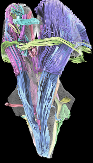
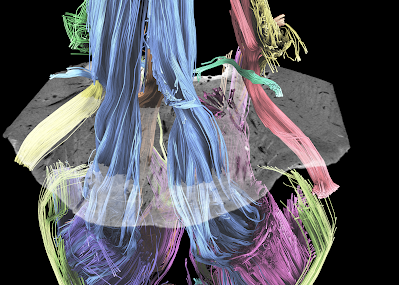
A HARDI scheme was used, and a total of 120 diffusion sampling directions were acquired. The b-value was 4000 s/mm2. The in-plane resolution was 0.2 mm. The slice thickness was 0.2 mm. The b-table was checked by an automatic quality control routine to ensure its accuracy (Schilling et al. MRI, 2019). The restricted diffusion was quantified using restricted diffusion imaging (Yeh et al., MRM, 77:603–612 (2017)). The diffusion data were reconstructed using generalized q-sampling imaging (Yeh et al., IEEE TMI, ;29(9):1626-35, 2010) with a diffusion sampling length ratio of 0.4.
Instructions
-
Windows: Download the civm_brainstem_dsi_studio_win64.zip file, unzip it, and run dsi_studio.exe (may needs to enable security setting).
-
Mac: Open on the dmg file (need to enable running third party package) and click on the DSI Studio icon to run it Memory requirement: > 16GB
The data, including the image files, trackable FIB.GZ file, and tractography files, are included in the program-data package. For Windows version, they are stored under the presentation folder of the zip file. For Mac, right click on the DSI Studio program to show content and navigator to /Contents/MacOS/presentation
License
2021 Center for In Vivo Microscopy, Duke University
- These data are made available as a service to the research community by Duke University through its Center for In Vivo Microscopy (“Duke CIVM”), located at DUMC-Center for InVivo Microscopy, Bryan Research Building, 311 Research Dr, Box 3302, Durham, NC 27710.
2.The data are provided for research and educational uses. No rights are provided to User under any patent applications, trade secrets, copyrights, or other proprietary rights of Duke CIVM, except as necessary to use the data and images for such research and educational purposes. Neither this data nor the results of use of the data shall be used as the basis for any commercial product or service offered to third parties- The user shall not transfer the data and images contained herein to any third party without express written permission from Duke CIVM. This shall not be taken to prevent the User from including representative images and data in academic publications of User’s research results.
- The User is encouraged to publish the results of his or her research. All publications based upon use of the data shall acknowledge the Duke University Center for In Vivo Microscopy and cite revelant publications.
Reference
1.Adil SM, Calabrese E, Charalambous LT, Cook JJ, Rahimpour S, Atik AF, Cofer GP, Parente BA, Johnson GA, Lad SP, White LE. A High-Resolution Interactive Atlas of the Human Brainstem Using Magnetic Resonance Imaging. NeuroImage. 2021 May 2:118135. 2.Calabrese, E., Hickey, P., Hulette, C., Zhang, J., Parente, B., Lad, S. P., & Johnson, G. A. (2015). Postmortem diffusion MRI of the human brainstem and thalamus for deep brain stimulator electrode localization. Hum Brain Mapp, 36(8), 3167–3178. https://doi.org/10.1002/hbm.22836


